
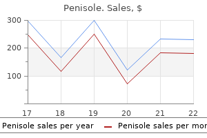
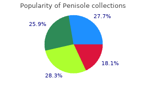
Penisole
Buy penisole overnight delivery
Measles (Rubeola) Documentation of the spread of infection through airborne droplets medications causing gout generic penisole 300 mg visa, contaminated water, or contact with inanimate Immunologic Disease objects is generally undocumented. Fusion of the viral Pemphigus Vulgaris membrane and the host cell membrane is mediated by the Mucous Membrane Pemphigoid sequential binding of cell surface viral glycoproteins to the Bullous Pemphigoid host cell membrane. During the primary infection, only a small percentage of individuals show clinical signs and symptoms of infectious systemic disease, whereas a vast majority experience only subclinical disease. The incubation period after Oral mucous membranes may be infected by one of several exposure ranges from several days to two weeks. In sympdiferent viruses, each producing a relatively distinct clinitomatic primary disease, a vesiculoulcerative eruption (prical-pathologic picture (Table 1-1). The large family herpesmary gingivostomatitis) occurs in the oral and perioral viridae (or herpesviruses) are structurally similar, charactertissues usually at the original site of contact. Seven types of herpesviruses lieved to migrate, through some unknown mechanism, are known to be pathogenic for humans, with six of these along the periaxonal sheath of the trigeminal nerve to the linked to diseases in the head and neck region. Both forms are self-limited in the immunocom(fever blisters), prior to a cold (cold sores), trauma, menstrual petent host, but recurrences of the secondary form are comcycle, stress, or immunosuppression causing a secondary or mon because the virus can be sequestered in a latent state recurrent infection. Control of symptoms rather than An immunocompromised host may develop severe seccure is the usual goal of treatment. Lesions caused by either and base of tongue virus are clinically indistinguishable. Viral shedding, a phenomenon in which a previously infected but asymptomatic individual may be capable of transmitting the virus, has been reported, although the relationship between frequency of shedding, viral titer, and actual transmission is unknown. Subclinical infection Reactivation Clinical Features Primary Herpetic Gingivostomatitis. This is in contrast to the recurrent form of the disease, in which lesions are confned to the lips, hard palate, and gingiva. Reactivation of latent herpes simplex virus type 1 Oral and perioral vesicles rupture, forming ulcers. Triggers: sunlight, stress, immunosuppression Intraoral lesions may be found on any surface. Reactivation common; frequency decreases with aging Systemic signs/symptoms include fever and malaise. Prodromal symptoms: tingling and burning Self-limited disorder; symptomatic care is provided. Clinical Features Affects perioral skin, lips, gingiva, palate Treatment Self-limited Acyclovir and analogs may control virus. Treatment Possible control with acyclovir and analogs Must be administered early Systemic treatment much more effective than topical treatment lesions are accompanied by fever, arthralgia, malaise, anDifferential Diagnosis orexia, headache, and cervical lymphadenopathy. Pemphigus vulgaris After the systemic primary infection runs its course of Erosive lichen planus Linear immunoglobulin (Ig)A disease about 7 to 10 days, lesions heal without scar formation. By Contact allergy this time, the virus may have migrated to the trigeminal Discoid lupus erythematosus ganglion to reside in a latent form. Regionally, most secondary lesions appear on It is believed that only rarely does reinfection from an exogthe vermilion and surrounding skin. Intraoral recurrences are physiology of recurrence has been related to a breakdown in almost always restricted to the hard palate or gingiva. Secondary herpes in the context of matory mediators that allows the virus to replicate. Patients immunosuppression results in signifcant pain and discomusually have prodromal symptoms of tingling, burning, or fort, as well as a predisposition to secondary bacterial and pain in the site at which lesions will appear. In contrast to those occurring in immunoof hours, multiple fragile and short-lived vesicles appear. Secondary by inoculation of the skin through a break in skin lesions typically occur at or near the same site with each integrity. In the case of a seronegative clinician, contact could result in a vesiculoulcerative eruption Pain, redness, and swelling are prominent with herpetic on the digit (rather than in the oral region), along with whitlow and can be very pronounced. Multiple lesions, neurologic prodrome keratinocytes cannot be demonstrated in biopsy or cyto(tingling), vesicles preceding ulcers, and palatal and gingival logic preparations. Primary herpetic gingivostomato herpetic lesions, aphthae are found almost exclusively on titis is usually apparent from clinical features.
Syndromes
- Feeling faint
- Respiratory failure
- Occurs frequently
- Some candles
- The pain is associated with medical problems, such as a history of herpes infections or a new rash
- Describe the amount of stool leakage (discharge with gas, a large amount of stool)?
Discount 300mg penisole with visa
Which criteria can be used to diagnose a basal cell Pili trianGuli anD CanaliCuli carcinoma A variable number of red medications multiple sclerosis purchase penisole 300mg without prescription, sharply demarcated vascular spaces called lacunae and fibrous septae 1. Pigment network, arborizing vessels, and central specific criteria such as symmetry of color and white patch structure and one prominent color. Usually they have several well-developed melanomaperiphery specific criteria such as asymmetry of color and C. A central white patch and peripheral pigment structure, multicomponent global pattern, regular network network, regular globules, and regular streaks. They contain a variable number of melanomalike cysts specific criteria such as asymmetry of color and strucE. Multifocal hypopigmentation, arborizing vessels, and ture, multicomponent global pattern, irregular local a central bluish white veil criteria, 5 or 6 colors, and polymorphous vessels. Melanoma-specific criteria on the trunk and extremities Answers can contain this combination of criteria: 1. Asymmetry of color and structure, a cobblestone variation of pigment network (regular and/or irregular), global pattern, and regular globules or blotches multiple brown dots and/or globules, homogeneous B. A multicomponent global pattern, symmetry of color blue color of a blue nevus, and parallel patterns seen on and structure, regular network, regular globules, and acral skin. The default category is the last way to diagregression nose a melanocytic lesion. Polymorphous vessels, arborizing vessels, 2 colors, openings can be seen in melanocytic lesions but are not and regular streaks primary criteria to make the diagnosis. Irregular network, irregular globules, irregular and C diagnose a basal cell carcinoma, dermatofbroma, blotches, and regression and hemangioma. Diagnosing a melanocytic lesion by default means that one does not see criteria for a melanocytic lesion, 8. Dysplastic nevi typically have the following combination seborrheic keratosis, basal cell carcinoma, dermatofof criteria: broma, or hemangioma. Symmetry of color and structure and no melanomaOne has to memorize all the criteria from each specifc specific criteria potential diagnosis to be able to diagnose a melanocytic B. Multifocal regression, peppering, regular pigment nique routinely in ones daily practice. Ink spot lentigo network, regular dots and globules and pyogenic granuloma are not in this algorithm. All the criteria used to diagnose seborrheic keratosis eral melanoma-specific criteria are commonly seen in daily practice. Asymmetry of color and structure plus several melacriteria can also be seen in atypical seborrheic keratosis. A Spitzoid lesion only refers to the starburst or pink but could be found in seborrheic keratosis. Melanoma is not in the differential diagnosis of reguand are not seen in seborrheic keratosis. In an adult, most Spitzoid lesions do not need to be and dermoscopy is used to confrm ones clinical impresexcised. Symmetrical and asymmetrical starburst patterns can nition, if one sees pigment network, the lesion could not be seen in melanoma. A subset of melanomas can be undistinguished from basal cell carcinoma with pigmen10. Which of the following statement best describes the critation and arborizing vessels. Moth-eaten borders are seen Tere are only 6 patterns (starburst, globular, in lentigines and fat seborrheic keratosis, never in basal homogeneous, pink, black network, atypical). Since symmetrical and asymmetrical Spitzoid spaces with well-demarcated sharp borders. Tere is no patterns can be found in melanoma, they should all be set number of lacunae needed to make the diagnoses. A dermatopaAt times one has to use their imagination to decide if thologist that specializes in melanocytic lesions is good, the margins ft the criteria for vascular spaces. Even experienced dermatopathologists have trouble Black homogeneous color usually represents thrombosis. Fibrous whitish and atypical Spitzoid lesions have the potential to metasseptae and/or bluish white color are routinely seen in tasize to regional lymph nodes and kill the patient. Superfcial spreading melanoma can have it all as far ferentiate lacunae and red color of a hemangioma from as the spectrum of melanoma-specifc criteria goes. The the milky red areas that can contain out-of-focus reddish criteria can be well developed or difcult to identify. The more high-risk criteria identifed in the in most cases dermoscopy is not needed to make the lesion, the greater the chance that one is dealing with a diagnosis. Nodular and amelanotic melanoma are more the primary criteria to make the diagnosis may or may likely to be feature poor or featureless. Tere are innumerable ways that the central white patch can appear, and in Argenziano G et al. Dermoscopy features of melanoma incognito: many cases it is not centrally located. Dermoscopy key points: recommendations from the that is pathognomonic for melanoma. Dysplastic nevi are ubiquitous in the light-skinned tologist part 1:dermoscopy of pediatric infectious and inflammapopulation and can be indistinguishable clinically and tory skin lesions and hair disorders. Dermoscopy for the pediatric dermatologist part 2: dermoscopy of genetic syndromes and cutaneous there are melanomas that do not have well-developed manifestations of pediatric vascular lesions. Dermoscopy for the pediatric derThey can have a variable number of melanomaspecifc matologist part 3: dermoscopy of melanocytic lesions. Identification of clinically featureless incipient melaMiteva M, Tosti A: Dermoscopy of hair shaft disorders. Atlas of Dermoscopy, multi-center study conducted by the international dermoscopy 2nd Ed. Chapter 32 Reflectance confocal MicRoscopy: the eRa of noninvasive iMaging Marigdalia K. Bright dots within keratinocytes correspond to inflammatory infiltrate (exocytosis).
Buy discount penisole 300mg on line
Hepatomegaly is a non patients bleed very easily and have hyperelastic specifc sign of many medical conditions but is skin medications vaginal dryness order penisole with a visa. Pain on urination account for a nephritic picture, but immuno would be a symptom of a urinary tract infec fuorescence would show an absence of any tion. Furthermore, if the patient in his urine, he most likely does not have a uri nary tract infection. It is possible that this seen in Parkinson disease and is unrelated to fetus inherited a recessive disorder such as cys rhabdomyolysis. These defects are caused meiosis is the usual cause of trisomy 21, the ge by the failure of the caudal portion of the neu netic abnormality in Down syndrome. On clinical neural tube defect include valproate and car examination, typical absence seizures appear bamazepine. Children are not responsive during (Potter syndrome) is caused by disruption in the seizure and are amnestic of what happened the interaction between the ureteric bud and during the attack. Clas does not produce urine (which is a component sically, a regular and symmetric 3-Hz spike is of amniotic fuid), there is a smaller volume of found on electroencephalography. This is described mide is the primary treatment option in cases by the term oligohydramnios. Carbamazepine has nary hypoplasia, fetal compression with altered been associated with the exacerbation of ab facies, and positioning defects of hands and sence seizures. These adjunctive therapies, however, is a connection between the pulmonary artery have limited effcacy. This pathway should associated with the exacerbation of absence be open during gestation and is not an abnor seizures. If the ductus arteriosus remains patent will be incorrectly attributed to inattentiveness after birth, the baby can be given indomethacin to help stimulate the vessel to close. They are abundant in cells that require a large amount of energy, such as myocytes. Secondary lysosomes multiple myeloma, a neoplastic proliferation are formed when a primary lysosome, with its of plasma cells in the bone marrow that often hydrolytic enzymes, fuses with materials for leads to lytic bone lesions and pathological degradation. Cells B lymphocyte that can produce and secrete such as macrophages, which are responsible large amounts of antibody specifc to a par for phagocytosis of cell debris, may contain ticular antigen. Areas of rough endo of one isotype and antigen specifcally, which plasmic reticulum in neurons are called Nissl can be detected as an M spike by serum pro bodies. A 41-year-old man is admitted to the hospital sician by her distraught parents because of a for progressive obtundation. The rest of the examination is worsens with the development of dysarthria, unremarkable. A 32-year-old woman presents to her family doctor complaining of fatigue, myalgia, and 4. Labo nation reveals cervical lymphadenopathy and ratory analysis reveals: the rash seen in the image. Colonoscopy of one of the af fected patients reveals colonic infamma tion with exudates and necrosis of the muco Courtesy of Dr. Which of the following is the micro biology laboratory likely to isolate from the af (A) Aortic aneurysm fected patients A 68-year-old woman presents to the emer (E) A gram-positive anaerobe gency department with altered mental status. A 9-year-old boy is brought to the emergency rate is 116/min, respiratory rate is 23/min, department with a two-day history of abdomi and blood pressure is 132/87 mm Hg. Which of the follow diffuse abdominal tenderness and a rash over ing is the most accurate description of the pa the arms and the legs. A renal biopsy specimen is obtained and reveals a focal proliferative glo merulonephritis, characterized by linear stain ing of the basement membrane on immuno fuorescence for IgG. He is subsequently found to have an eye tumor that is caused by dysfunction of a spe B cifc cell-cycle regulatory gene product. A biopsy is taken, and fever, diaphoresis, mydriasis, and severe agita the results are shown in the image. When asked, his mother states that her the following is the most likely diagnosis A 60-year-old woman is receiving chemother sistently localized to the right lower quadrant apy for breast cancer. She presents to her on without migration, and has been associated cologist complaining of fatigue and dyspnea with nausea and vomiting. Physical examination reveals an ods are usually regular, her last menstruation elevated jugular venous pressure, crackles, and was approximately six weeks ago. X-ray of the chest tion, she is found to be afebrile with a blood shows an enlarged cardiac silhouette. Her on pressure of 90/60 mm Hg, a pulse of 110/min, cologist believes her chemotherapeutic agent and a respiratory rate of 26/min. What is the examination shows localized tenderness with mechanism of the chemotherapeutic agent she guarding in the right lower quadrant. Laboratory stud (B) Inhibition of dihydrofolate reductase ies show a hematocrit of 29. A 22-year-old woman comes to your offce Glucose: 96 mg/dL complaining of vaginal itching and burn Serum osmolality: 250 mOsm/kg ing. Vaginal examina tion reveals the lesion seen in the image, and (A) Excessive fuid intake treatment is started. The patient subsequently (B) Glioblastoma multiforme develops elevated levels of blood urea nitro (C) Metastatic breast cancer gen and creatinine in addition to a tremor and (D) Small cell lung cancer mental status changes. Which of the following (E) Squamous cell lung cancer agents did this patient most likely receive A 33-year-old man from upstate New York comes to his physician because of fu-like symptoms after a camping trip one week ago. He also notes a troubling rash on his leg that has moved slowly from his ankle to his midthigh over the past several days. The patient recently lost his pre scription benefts through his health insurance plan, so he uses an old bottle of tetracycline from his medicine cabinet at home. One week later the man presents to the emergency de partment with signs of dehydration and a cre atinine level of 3. When asked about into the emergency department after having a her sleeping habits in greater depth, she re witnessed tonic-clonic seizure lasting two min ports sleeping for seven-nine hours per night, utes. Laboratory gressively more animated and begins making tests show: aggressive sexual advances toward the physi cian and the staff. She reports that her mood Na+: 123 mEq/L has been good, but not overly elevated, expan K+: 3. The following graph is a depiction of the Star (E) Schizotypal personality disorder ling curve showing the relationship between cardiac output and ventricular end diastolic 21.
Purchase penisole cheap
Decreased intake medicine 122 300 mg penisole amex, however, is not the with hypertonic saline, but rather the opposite, pathogenesis of his disease. Diffuse axonal injury teodystrophy can have normal or even elevated occurs in the setting of central nervous system vitamin D intake. Cholecalciferol, or vita sults in disruption of the axon at the nodes of min D3, is found in meats. Diffuse axonal injury does not occur in humans in the dermis by ultraviolet light with electrolyte abnormalities. Intracerebral hemor can have normal or even elevated vitamin D rhage can occur as a result of hypertension, intake. The description of it does not occur as a result of hyponatremia or colonic infammation with exudates and ne the associated treatment. Uncal herniation can fcile, of which there have been several out result only from focal processes within the cra breaks. C diffcile is a gram-positive anaerobe nial vault, such as intracranial hemorrhage, spore-former that produces toxin A (which but does not occur with diffuse processes asso causes diarrhea) and toxin B (which is cy ciated with electrolyte abnormalities. This man has renal morbidity and even mortality associated with osteodystrophy, a common complication of C diffcile colitis. In these patients, decreased conversion of 25-hydroxyvitamin Answer A is incorrect. Salmonella is an exam D to the active 1, 25-dihydroxycholecalciferol ple of a gram-negative facultative intracellu in kidney cells leads to decreased calcium ab lar organism and could produce diarrhea, but sorption and thus a low serum calcium level. Gram-negative lactose to counteract the low calcium levels by in fermenters that can cause diarrhea include creasing bone resorption. Gram-negative lactose with calcium, phosphate binders, and calcitriol nonfermenters that can cause diarrhea include (synthetic vitamin D) supplementation. Her bicarbonate level is low, so this is a is the most common small-vessel vasculitis in metabolic acidosis. The disease has appropriate respiratory compensation us is self limiting and treatment is supportive. The 3 tomatology is more likely to have a vasculitis expected partial pressure of carbon dioxide associated with antineutrophil cytoplasmic an would therefore be 16. IgA antibody deposi simultaneous respiratory alkalosis that is more tion in the mesangium is the characteristic than compensatory. This patient is acidotic, low-grade proteinuria during or a few days after and her bicarbonate level is low, so we know an infection. IgG antibodies against platelets is the pathophysiology of idiopathic Answer E is incorrect. The patient is experi thrombocytopenic purpura, which is charac encing a respiratory alkalosis, but there is also terized by thrombocytopenia leading to mu a simultaneous metabolic acidosis. IgG antibodies depos in basement membranes in the lung and kid ited in the glomerular basement membrane is ney, leading to nephritic kidney disease and the etiology of Goodpasture syndrome, which a restrictive lung disease. The patient is experi sifed as pathologies that result in decreased encing a metabolic acidosis, but there is also a lung volumes with a relative preservation of simultaneous respiratory alkalosis. Labetalol is a non strictive pattern seen because of restriction of selective a and b-antagonist that blocks many lung expansion. Atropine is a musca taglandins are produced in response to in rinic antagonist that would be appropriate creased sympathetic activity and act to pref therapy for overdose of an acetylcholinesterase erentially vasodilate afferent arterioles. Flumazenil is a ben vasoconstriction of the efferent arterioles and zodiazepine receptor antagonist. The clini son that drugs blocking the formation of pros cal features of acute benzodiazepine intoxica taglandins (ie, nonsteroidal anti-infammatory tion include slurred speech, lack of coordina drugs) are damaging to the kidney is that they tion, unsteady gait, and impaired attention or block vasodilation at the afferent arterioles and memory. Fluoxetine is a selec cause vasoconstriction of B, while the prosta tive serotonin reuptake inhibitor. In inherited cases, the frst receptor antagonist that would be appropriate hit is carried in the germline. A second hit to therapy for an opiate overdose such as with any retinoblast will result in cancer, making it heroin or morphine. In with opioid overdose would appear sleepy, le sporadic cases, both hits have to occur in the thargic, or comatose, depending on the degree same retinoblast. Pupils would be miotic, not myd tumors are typically solitary and more often oc riatic. The G2/M check acetylcholinesterase inhibitor that might be point is another important cell-cycle regula used for an antimuscarinic drug overdose, tory checkpoint. It provides another oppor such as with atropine, scopolamine, or Jimson tunity to prevent the cell from undergoing weed. In addi toma may present in one eye, as in this patient, tion, it also initiates the apoptotic cascade in or bilaterally, as in approximately 30% of cases. The Rb protein does toma is called sporadic, in contrast with the fa not promote histone acetylation. In fact, the milial form, which is associated with a family Rb-E2F complex promotes histone deacety history. The patient has a priate implantation of a blastocyst in a normal likely diagnosis of basal cell carcinoma. It is pregnancy and is therefore not directly associ characterized by pink or fesh-colored pearly ated with pathology. This describes the ules are locally invasive but usually non likely etiology of acute appendicitis. There is a purely basaloid population with periumbilical pain that later migrates to the minimal stromal response. Although this can be associated with premalignant lesion characterized by small, irregular bleeding and abdominal/pelvic pain, rough erythematous or brownish papules. Dermatitis herpetifor tilize a single ovum, a partial hydatidiform mis is a dermatologic condition associated with mole is formed. Like a ruptured ectopic preg celiac disease that is characterized by pruritic nancy, this will produce vaginal bleeding and papules and vesicles. The a benign, fat, pigmented squamous prolifera occipital pole is the extreme posterior end of tion with keratin cysts. The macula is re noma commonly appears on the hands and sponsible for central vision. Methotrexate is common cause of optic chiasm lesions is a pi also used as an anti-infammatory agent and to tuitary adenoma. These agents are not associated clusion with sparing of the vessels supplying with cardiotoxicity. Adverse effects include endometrial macular sparing seen in our patient, a phe hyperplasia and carcinoma, increased bone nomenon caused by sparing of the occipital density, hypercoagulability, hot fashes, night pole. Neurotoxicity can mani use of these agents can lead to congestive heart fest as lethargy, confusion, tremor, delirium, failure, as seen in this patient, particularly a or seizures. Other adverse effects of a crystalluria that can lead to obstructive of doxorubicin include bone marrow suppres nephropathy or interstitial nephritis. Patients fuorouracil, busulfan, cisplatin, mitoxantrone, complain of pain, itching, dysuria, and vaginal and paclitaxel. Fluconazole is an an and headache, and the aerosolized form is as tifungal agent that inhibits fungal steroid syn sociated with bronchospasm, rash, and con thesis. The discharge classically associated ary to an extensive history of smoking, and the with vaginal candidiasis is thick and white. Infection dition, a patient who is purely drinking too with T vaginalis presents with a malodorous, much should have relatively appropriate com frothy green discharge, vaginal pruritus, and pensatory mechanisms, such as the ability to erythema. This would lead to ally due to Gardnerella vaginalis infection, water loss from the kidneys to compensate, and presents with perivaginal infammation and thus serum osmolality should not decrease.

Cheap penisole 300 mg free shipping
Non-steroid anti-inflammatory drugs (ibuprofen) or acetaminophen were recommended in case of injection site reaction but were not routinely administered by protocol at any time during the study treatment zenkers diverticulum purchase genuine penisole. Re-initiation of therapy with Rebif following elevation of liver function tests could only be considered once. In addition, at the discretion of the Investigator, corticosteroids could be either stopped abruptly or tapered over a maximum of 10 days. Patients were not required to discontinue the treatment period solely based on the occurrence of a relapse, unless the patient or Investigator determined that he or she had met the criteria for withdrawal. Reviewer Comment: the frequency that potential relapses were evaluated within 7 days of onset is assessed in the following tables: (Table 25, Table 26, and Table 27). For each relapse that satisfied the 3 criteria above, it was then determined whether the potential relapse was within 30 days. If a potential relapse was within 30 days, then the potential relapse was not a protocol-defined relapse. Study Endpoints Primary Efficacy Endpoint the primary efficacy endpoint was the annualized protocol-defined relapse rate at two years (96 weeks). Patients who received an incorrect therapy from that which was intended are included in the efficacy analyses according to their randomized treatment. The annualized relapse rates by 96 weeks are analyzed using a negative binomial model. The proportion of patients with confirmed disability progression was estimated using Kaplan-Meier methodology. The overall hazard ratio was estimated using a stratified Cox regression model with the same stratification factors used in the stratified log-rank test above. Data from other unscheduled assessments are not included in this summary or analysis. A negative binomial model is used to compare the difference between ocrelizumab and Rebif groups. The Total Number of New, and/or Enlarging T2 Hyperintense Lesions as Detected by Brain Magnetic Resonance Imaging at Week 24, Week 48 and Week 96 the same approach has been used for the statistical analysis of new and/or enlarging T2 hyperintense lesions as for the total number of T1 Gd-enhanced lesions. The same approach to data derivation is used for disability improvement as for disability progression. All patients without disability improvement will be counted as not improved, independent of follow-up time. Data from the two studies with respect to ocrelizumab group vs Rebif group will be pooled for analysis of this endpoint. Time to confirmed disability progression (24-week confirmation) is defined as the time from Baseline (Day 1) to the first disability progression, which is confirmed at the next regularly scheduled visit 161 days after the initial disability progression. All initial disability progression events up to Week 96 with corresponding confirmation visits at the next scheduled visit are taken into account for the statistical analysis. Data from the two studies with respect to ocrelizumab group versus Rebif group have been pooled for analysis of this endpoint. Total Number of T1-Hypo-Intense Lesions (Chronic Black Holes) at Weeks 24, 48, and 96 the same approach has been used for the statistical analysis of T1 hypointense lesions as for the total number of T1 Gadolinium-enhanced lesions. Patients who discontinued treatment early with at least one event before early discontinuation were considered as having evidence of disease activity. Even if an event was not reported before early discontinuation, the patient was considered as having evidence of disease activity if the reason for early discontinuation is lack of efficacy or death; otherwise, it was considered a missing observation. Any potential impact of disclosed financial interest on overall efficacy or safety outcomes is therefore expected to be limited. Reviewer Comment: There were relatively few investigators who reported a disclosable financial interest and there were relatively few patients enrolled at these sites. Patient Disposition First patient randomized: 31 August 2011 Last patient randomized: 14 February 2013 Data cut-off date: 2 April 2015 1041 patients were screened and 821 were enrolled and randomized. Two of these were due to subject withdrawal of consent, one due to physician decision and one due to a protocol violation. Study Results Compliance with Good Clinical Practices See page 51 Financial Disclosure See page 51 Patient Disposition First patient randomized: 20 September 2011 Last patient randomized: 28 March 2013 Data cut-off date: 12 May 2015 835 patients were enrolled at 166 sites in 24 countries. The safety population therefore consisted of 834 patients treated, 417 in each treatment group. The study consisted of a screening period, a blinded treatment period, an open label treatment period and a safety follow-up period. Following informed consent subjects entered a screening period of up to 8 weeks to determine eligibility. Blinded treatment was continued until the study was considered completed and treatment unblnded. This was to occur when the last enrolled subject completed at least 120 weeks of treatment. However if the target number of 12 week confirmed progressions had not occurred at that point then the blinded treatment period would be extended until at least 253 confirmed progressions occurred. Reviewer Comment: Note that reconsent was required for those patients who reached the 24 Week Confirmed Disability Progression endpoint. In addition, a structured telephone interview was conducted on a 4-week basis between study visits from Week 8 through the end of the Blinded Treatment Period to identify any new or worsening neurological symptoms that could warrant an unscheduled visit. Additional unscheduled visits for the assessment of potential relapses, new neurological symptoms or safety events could occur at any time. If patients had received corticosteroids for a relapse, the scan should have been done prior to the first steroid dose if the pre-steroid scan was within 1 week of the scheduled visit. For patients who have received corticosteroids, there should have been an interval of 3 weeks between the last dose of corticosteroids and the scan. The centralized reading center was blinded to the treatment assignment and the reading was performed in the absence of clinical information. Following unblinding subjects were eligible to enter an open label treatment period if the investigator determined that the subject could benefit from continued (for those who were on ocrelizumab) or initiation of treatment with ocrelizumab. Patients who discontinued treatment for any reason were to be followed up for at least 48 weeks after the last infusion in the Safety Follow-Up Period with visits every 12 weeks until 48 weeks had elapsed since the last infusion of study drug. The full schedule of assessments during the blinded treatment phase is shown in Table 99. Symptoms must have persisted for more than 24 hours and could not be attributable to confounding clinical factors such as a fever, infection, injury, or adverse reactions to concomitant medications. Episodic spasms, sexual dysfunction, fatigue, mood change or bladder or bowel urgency or incontinence did not suffice to establish a relapse. It should be noted that all patients with new neurological symptoms defined at a visit or over the phone should have been referred to the Examining Investigator unless the Treating Investigator considers the symptoms consistent with an intensification of neurological symptoms from a transient systemic infection. The following laboratory results were provided to the Treating Investigator because they were Criteria for Retreatment with Ocrelizumab (See Section 6. Confirmation of disability progression was required at a regularly scheduled visit that was at least 12 weeks after the initial disease progression. An interim data lock was to occur when the last patient had completed the Week 120 assessment. If additional treatment cycles were instituted due to lower than anticipated disease progression rates at 120 weeks, then the interim data lock was to occur when approximately 253 sustained disability progression events had occurred. An additional analysis comprising of both safety and efficacy endpoints was planned at the end of the follow-up period to investigate the maintenance of the treatment effect and/or the potential for a withdrawal effect. An assessment that occurred within 30 days after a protocol-defined relapse was not to be used for confirmation of sustained disability progression.
Tormentillae rhizoma (Tormentil). Penisole.
- What is Tormentil?
- Bleeding, fever, stomach complaints, diarrhea, and mild swelling (inflammation) of the mouth and throat.
- Dosing considerations for Tormentil.
- How does Tormentil work?
- Are there safety concerns?
Source: http://www.rxlist.com/script/main/art.asp?articlekey=96372
Generic penisole 300mg on-line
This variant Adenosquamous and squamous carcinocases of undifferentiated lymphoepitheresembles the tumour arising in salivary ma symptoms 16 dpo cheap 300 mg penisole visa. A predominant component of extracellular mucus (mucus lakes), usually visible to the naked eye, is present in the stroma. A malignant tumour in which there is a predominance of discrete cells distended with mucus. A cholangiocarcinoma with spindle cell areas resembling spindle cell sarcoma or fibrosarcoma or with features of malignant fibrous histiocytoma. B Cytokeratin 7 immunohistochemistry demonstrates tumour cells spreading along bile ducts and infiltrating liver tissue. The hepatocyte antily, and poorly differentiated adenocarcigoblet cell metaplasia {2008, 913}. In hepatolithiasis, the findings static adenocarcinoma of biliary tract or lary structures, moderately differentiated are those of cholangitis, with proliferation pancreatic origin. Occasionally, dysplastumours show moderately distorted tubuof the biliary epithelial lining and peritic changes in neighbouring bile ducts lar patterns with cribriform formations biliary glandular cells, and multiple foci suggest intrahepatic origin. In addition, and/or a cord-like pattern, while the poorof biliary intraepithelial neoplasia . Some peribiliary Dilated intrahepatic and extrahepatic bile glands may also be dysplastic. They are usually multiple and 178 Tumours of the liver and intrahepatic bile ducts A. Within a biliary anomalies, and also in normal livnosis of a well-differentiated cholangiofibrous or hyalinized stroma, they present ers, multiple cysts may be seen around carcinoma. It occurs in apparently noras irregular or round ductal structures the intrahepatic large bile ducts {1319, mal livers and also in acquired liver disthat appear somewhat dilated and have 1320}. These cysts are derived from peribillumina contain proteinaceous or bileiary glands and should be differentiated Molecular genetics and genetic susstained secretion. Some ducts may be cystiand Korean patients show an intermediducts with cuboidal cells that have regucally dilated. Their fibrous stroma shows varying degrees of chronic inflammation and collagenization. Biliary adenofibroma is characterized by a complex tubulocystic biliary epithelium without mucin production, together with abundant fibroblastic stromal components . Its expansive growth, and foci of epithelial tufting, cellular atypia and mitoses favor a neoplastic process. Small, normal appearing proliferating bile ducts associated with a small conIn chronic advanced liver disease and nective tissue component and lymphocytic infiltration. Biliary gesting a somewhat more favourable mutations and less frequently nonsense epithelial cells are continuously exposed prognosis . One study found the ly detectable as a small nodule during folOverexpression of c-erbB-2 occurs in one 5-year survival rate was 39% in patients low-up of hepatitis virus-related cirrhosis, fourth to about two thirds of carcinoma of with mass-forming tumours and 69% for and is treatable with hepatectomy . Ponchon and cholangiocarcinoma Definition Epidemiology noexpression is sufficient for the diagnoA rare tumour containing unequivocal this tumour type comprises less than 1% sis of a hepatocellular carcinomatous elements of both hepatocellular and of all liver carcinomas. Gross inspection does not show significantly different morphology compared to hepatocellular carcinoma. In tumours with a major cholangiocarcinomatous component with fibrous stroma, the cut surface is firm. Histopathology Combined hepatocellular and cholangiocarcinoma is the term preferred for a A tumour containing both hepatocellular A and distinct or separate cholangiocarcinoma. This category should not be used for tumours in which either form of growth is insufficiently differentiated for positive identification. Hepatocytes preferentially express cytokeratins 8 and 18 and, like duct epithelial B cells, cytokeratins 7 and 19. Ponchon and cystadenocarcinoma Definition solid areas of grey-white tumour in a females and has been likened to ovarian A cystic tumour either benign (cystadethickened wall. The stromal cells express noma) or malignant (cystadenocarcinovimentin, and there are many cells that ma), lined by epithelium with papillary Tumour spread and staging express smooth muscle actin. A xaninfoldings that may be mucus-secreting Cystadenocarcinomas show intrahepatic or, less frequently, serous. Lesions arise spread and metastasis to regional lymph from ducts proximal to the hilum of the nodes in the hepatoduodenal ligament. They differ from tumours that arise Distant metastases occur most frequent in in cystic congenital malformation and in the lungs, the pleura and the peritoneum. Epidemiology Bile duct cystadenoma and cystadenoHistopathology carcinoma are rare . Cystadenoma Cystadenomas are usually multilocular is seen almost exclusively in females, and are well defined by a fibrous capwith cystadenocarcinoma appearing sule, which may contain smooth muscle equally in males and females. Patients often present with abdominal the mucinous type is more common and pain and mass. Imaging resting on a basement membrane; polytechniques show multilocular cystic poid or papillary projections may be prestumour(s), occasionally with tiny papillary ent. Subjacent to the baseThe cysts are usually multilocular and ment membrane is a cellular, compacted typically range from 5 to 15 cm diameter mesenchymal stroma, which in turn is. This intrahepatic large bile duct in a case of hepatolithipapillary mass may occur as well as mesenchymal component is seen only in asis. A Large peribiliary cysts in the connective tissue of the hilus; the background liver shows advanced cirrhosis. Differenticells, cholesterol clefts and pigmented ation from intrahepatic bile duct cystadelipofuscin-containing macrophages, may noma depends on the demonstration of be present in the cyst wall. The serous cytological (particularly nuclear) atypia, type consists of multiple, small locules mitosis, and invasion of the underlying lined by a single layer of cuboidal cells stroma. The cells rest on a basement memmay be misdiagnosed as bile duct cysbrane but are not surrounded by the tadenomas because insufficient sammesenchymal stroma typical of the mucipling results in tumour morphology nous variety. Malignant change may not involve all of the Prognostic factors epithelium lining the cyst; it is usually mulThe prognosis of patients with biliary course of patients with unresectable tifocal. The tumours are so well defined duct cystadenocarcinomas is good if a tumours seems to be better than of that complete removal can usually be curative resection is possible. A malignant embryonal tumour with Less frequently nausea, vomiting, and divergent patterns of differentiation, abdominal pain are present. Rarely, tumour cells thelial hepatocytes, to embryonal cells, may produce human chorionic gonaand differentiated tissues including dotrophin, leading to precocious puberty osteoid-like material, fibrous connective with pubic hair, genital enlargement and tissue and striated muscle fibers. Epidemiology Hepatoblastoma is accompanied by aneHepatoblastoma is the most frequent mia in 70% of cases and by thrombocy. Only 3% are seen in ed in about 90% of patients at the time of patients over 15 years of age. Other laboratory abnormalities can Macroscopy Multiple masses, seen in the other 20% include elevated levels of serum cholesHepatoblastomas vary in size from 5 to of cases, may occur in either or both terol, bilirubin, alkaline phosphatase, and 22 cm in diameter and from 150 to 1, 400 lobes. Areas of necrosis and haemorrhage are usually present and Birth to Birth to may appear as soft or gelatinous, brown 2 years (%) 20 years (%) Type of Tumour (285 cases) (716 cases) to red tissue . These cells contain a by more abundant cytoplasm and larger small round nucleus with fine nuclear nuclei. Canaonly those cases in which macrotrabeculiculi may be seen between hepatocytes lae are a prominent feature of the lesion. In biopsies large, well demarcated lesion with central haemortaken before preoperative chemotherarhage. Hepatoblastomas also spread to the focal staining of the sinusoidal Beckwith-Wiedemann syndrome bone, brain, ovaries, and the eye {179, endothelial cells of normal liver . The cells display little cohesivedysplasia, absence of portal vein melanin pigment. While often interHypoglycemia resembling the hepatocytes of the develmixed with the fetal epithelial cells, the Inguinal hernia foci of embryonal cells, which are devoid Isosexual precocity of glycogen and lipid, can be identified Maternal clomiphene citrate and Pergonal Table 8.
Discount penisole master card
Exact pathogenesis of alcoholic liver predisposed to alcoholic cirrhosis is not clearly known medications for osteoporosis order line penisole, but injury is yet unclear as to why only some chronic alcoholics a few risk factors have been implicated. These are as under: develop the complete sequence of changes in the liver while 1. However, knowledge and understanding of attributed alcoholic cirrhosis to chronic alcoholism. Available the ethanol metabolism has resulted in discarding the old evidence suggests that chronic and excessive consumption concept of liver injury due to malnutrition. Instead, now it of alcohol invariably leads to fatty liver in >90% of chronic is known that ethanol and its metabolites are responsible for alcoholics, progression to alcoholic hepatitis in 10-20% cases, ill-effects on the liver in a susceptible chronic alcoholic having and eventually to alcoholic cirrhosis in more than 10 years. Briefly, the biomedical and It is generally agreed that continued daily imbibing of 60-80 cellular pathogenesis due to chronic alcohol consumption gm of ethanol in any type of alcoholic beverage for at least culminating in morphologic lesions of alcoholic steatosis 10 years is likely to result in alcoholic cirrhosis. Liver injury (fatty liver), alcoholic hepatitis and alcoholic cirrhosis can is related to the quantity of ethanol contained in alcoholic be explained as under and is schematically illustrated in beverage consumed and its duration, but not related to the. There is evidence to alcoholic beverage is given on the label of the container, but suggest that ethanol ingestion for a period of 8-10 days regu in general, it s about 4-6% in beer, 10-12% in wine, and about larly may cause direct hepatotoxic effect on the liver and 40-50% in brandy, whisky and scotch. Ethanol is directly toxic to micro for long duration is less harmful since the liver is given tubules, mitochondria and membrane of hepatocytes. Women have increased susceptibility to develop hepatotoxic effects of ethanol are exerted by its metabolites, advanced alcoholic liver disease with much lesser alcohol chiefly acetaldehyde. Acetaldehyde produces progression is unclear but is probably linked to effects of hepatotoxicity by production of two adducts: oestrogen. Absolute or relative malnutrition of toxic and can cause cytoskeletal and membrane damage and proteins and vitamins is regarded as a contributory factor in bring about hepatocellular necrosis. Theses adducts have also a role in hepatic derived from alcohol displace other nutrients leading to fibrogenesis due to peroxisome proliferator-activated malnutrition and deficiency of vitamins in alcoholics. Additional factors contributing to malnutrition in alcoholics are chronic gastritis and pancreatitis. Cell-mediated immunity is in cirrhotic patients and may accelerate the course of the impaired in alcoholic liver disease. Ethanol causes direct occurrence of gout, impaired gluconeogenesis and altered 621 steroid metabolism. Alcohol is inhibitory to secretion of newly-synthesised proteins by the liver leading to their retention in the hepatocytes. Water is simultaneously retained in the cell in proportion to the protein and results in swelling of hepatocytes resulting in hepatomegaly in alcoholics. Chronic ingestion of alcohol results in increased oxygen demand by the liver resulting in a hypoxic state which causes hepatocellular necrosis in centrilobular zone (zone 3). In chronic alcoholism, there is rise in the amount of fat available to the liver which could be from exogenous (dietary) sources, excess mobilisation from adipose tissue or increased lipid synthesis by the liver itself. In a proportion of cases, Grossly, the liver is enlarged, yellow, greasy and firm with alcohol-related liver cell injury continues unabated despite a smooth and glistening capsule. Chronic ethanol ingestion is not only lymphocytes, macrophages and some multinucleate giant injurious to hepatocytes but also damages the intestinal cells. The injured intestinal cells elaborate endotoxins which release proinflammatory cytokines, chiefly tumour necrosis 2. These cytokines and develops acutely, usually following a bout of heavy drin endotoxinaemia produce apoptosis and necrosis of king. Repeated episodes of alcoholic hepatitis super hepatocytes and initiate inflammatory reaction in the alcohol imposed on pre-existing fatty liver are almost certainly a damaged liver. Mallory bodies are highly redox potential has been implicated in a number of metabolic suggestive of, but not specific for, alcoholic hepatitis since consequences such as in fatty liver, collagen formation, 622 Figure 21. Most of the hepatocytes are distended with large lipid vacuoles with peripherally displaced nuclei. In the liver being large, fatty and weighing usually above more extensive necrosis, the inflammatory infiltrate is 2 kg (. Eventually over a span of years, the liver more widespread and may involve the entire lobule. This nodules of the liver due to their fat content are tawny is also termed as creeping collagenosis. On cut section, spheroidal or angular from central vein to portal regions, or portal tract to portal nodules of fibrous septa are seen. As the fibrous scarring increases with time, Microscopically, alcoholic cirrhosis is a progressive the fibrous septa become dense and more confluent. Its features include the following iii) Hepatic parenchyma: the hepatocytes in the islands (. It shows nearly uniform-sized micronodules, devoid of central veins and having thick fibrous septa dividing them. There is minimal inflammation and some reactive bile duct proliferation in the septa. The hepatic parenchyma within the Post-necrotic cirrhosis, also termed post-hepatitic cirrhosis, nodules shows extensive fatty change early in the disease. Thus, there is an inverse bands of connective tissue and occurring most commonly relationship between the amount of fat and the amount after previous viral hepatitis. Based on epidemiologic and serologic studies, iv) Necrosis, inflammation and bile duct proliferation: the following factors have been implicated in the etiology of the etiologic clue to diagnosis in the form of Mallory post-necrotic cirrhosis. The fibrous septa usually contain sparse recent or remote attacks of acute viral hepatitis followed by infiltrate of mononuclear cells with some bile duct chronic viral hepatitis. Bile stasis and increased cytoplasmic hepatitis B and C; hepatitis A is not known to evolve into haemosiderin deposits due to enhanced iron absorption cirrhosis. The laboratory findings in the course of alcoholic of cases may have origin from toxicity due to chemicals and liver disease may be quite variable and liver biopsy is drugs such as phosphorus, carbon tetrachloride, mushroom necessary in doubtful cases. Prolonged prothrombin time and partial thromboplastin cirrhosis is macronodular type. Neutrophilic leucocytosis in alcoholic hepatitis and in scars and nodules of varying size (. Fibrous septa dividing the hepatic parenchyma into nodules are thick and contain prominent mononuclear inflammatory cell infiltrate and bile ductular hyperplasia. Biliary Cirrhosis surface shows scars and nodules varying in diameter from 3 mm to a few centimeters. Biliary cirrhosis is defined as a chronic disorder characterised Microscopically, the features are as follows (. Nodular pattern: the normal lobular architecture of continued cholestasis of intrahepatic or extrahepatic origin. However, Primary biliary cirrhosis in which the destructive process uninvolved portal tracts and central veins in the hepatic of unknown etiology affects intrahepatic bile ducts. Fibrous septa: the fibrous septa dividing the variable Primary sclerosing cholangitis and autoimmune cholangiopathy causing biliary cirrhosis. The etiology of these forms of biliary cirrhosis Active liver cell necrosis is usually inconspicuous. The etiology of this type infiltrate which may even form follicles, especially in cases remains unknown. Often there is extensive implicated: proliferation of bile ductules derived from collapsed liver 1. However, presently the most widely accepted hypothesis described on page 630, post-necrotic cirrhosis is seen as is autoimmune origin of the disease. In support are the frequent in women as in men, especially in the younger age following observations: group. Like in alcoholic cirrhosis, the patients may remain increased incidence of associated autoimmune diseases asymptomatic or may present with prominent signs and.
Cheap penisole online visa
The third Bethesda conference held from April 30 to May 2 treatment jaundice proven penisole 300mg, 2001 further revised the terminology. The Bethesda System has enjoyed widespread popularity, and it has been adopted almost worldwide to replace the outdated Papanicolaou numerical class system for reporting Pap smears. At the present time the ThinPrep Pap Tests has replaced nearly 100% of the cervical screening market in the United States. The morphology of squamous and glandular cells is preserved but the cellular patterns or arrangements associated with some lesions are altered, requiring slight modifications of cytodiagnostic criteria. Attaining proficiency in the interpretation of liquid based cytology will require a lot of time and effort from the cytology community worldwide. These new technologies have increased the cost of medical care, and at this moment they are not considered to be cost effective. Prior to 2002 various organizations generally recommended that screening should start at the beginning of sexual activity. It should continue annually throughout life, and women with several prior negative Pap smears may have the test repeated at longer intervals. The revised cervical cancer screening guidelines of the American Cancer Society and those of the American College of Obstetricians and Gynecologists were published in 2002 and 2003. Their recommended screening starts between the ages of 20 and 25 years and continues every 3 to 5 years until age 60 to 65. Many developing countries do not currently have any cervical cancer screening programs. Therefore, women should continue to have cervical cancer screening by Pap smear regardless of their vaccination status. It encompasses laboratory procedures, diagnostic accuracy and reliability with the goal to reduce false-positive and false-negative results. Results of re-examination of cytologic materials are statistically documented annually for evaluation of the laboratory performance. In 1943, Cary was the first investigator to report on the use of a metal canula and a syringe to aspirate endometrial cells. This sampling technique was never adopted because of the fear of seeding endometrial cancer cells into the peritoneal cavity. Butler and associates subsequently modified the Ayre brush and the technique for removing endometrial cells from the bristles of the brush. Despite a high diagnostic accuracy for endometrial carcinoma, the brushing technique was not widely 12 adopted. In 1964, Dowling and Gravlee reported on the combination of endometrial aspiration with uterine lavage under negative pressure to obtain endometrial cells. This technique did not produce consistently acceptable results because of the poor preservation of cell morphology, and it was subsequently discontinued. This sampling technique was widely used and achieved a high diagnostic accuracy rate for endometrial cancer. In 1973, Milan and Markley reported on the use of a plastic helix to sample the endometrium by scraping. In recent years, a few endometrial scraping devices have been introduced to clinical practice. They are simple to use and cause little or no discomfort to patients and the cell samples obtained have a low rate of cellular inadequacy. In 1971, Kjellgren and coworkers utilized this technique to classify ovarian cancers and have published the results in their 1972, 1974 and 1979 publications. Cell samples from the uterine cervix and posterior vaginal fornix are usually collected, respectively, by the pointed end and the blunt end of an Ayre spatula. They are either deposited onto 2 glass slides or mixed together on one glass slide. The use of a cytobrush in conjunction with a spatula can help ensure an adequate representation of the squamocolumnar junction. Once the samples have been evenly spread on the glass slide, they are immediately fixed with a commercial spray fixative. To prevent the formation of thick cellular ridges, the slide should be held about 25 cm from the spray nozzle or floated within the fixative for 15 to 30 min. The slides are then air-dried and placed in a rigid 13 container for mailing to a referral cytology laboratory. If the slide is broken during transportation, the cytologic material may be transferred to a new glass slide for examination by using a special technique. The ThinPrep Pap Test (Hologic Inc) consists of rinsing the collection device into a vial of a methanol-based fixative media (PreservCyt). After gentle dispersion to homogenize the specimen, the solution is aspirated through a membranous filter which is subsequently pressed against a glass slide. Negative pressure and surface tension allow for transfer of the cells to the slide, which is then automatically placed in 95% ethanol for fixing before staining. The automated processor homogenizes the specimen and dispenses it onto a density gradient reagent where cells are separated from interfering cell debris and inflammatory cells. Automated pipetting transfers the cell concentrate to small plastic chambers for gravity sedimentation onto glass slides and then subsequent Pap staining within each chamber. Any minute tissue fragments obtained are fixed in 10% neutral buffered formalin for supplementary histological examination. The cell films are either wet-fixed with 95% ethanol for staining with the Papanicolaou technique or hematoxylin and eosin, or air-dried for staining by the May-Grnwald-Giemsa technique or by one of its modified methods (Giemsa and Diff-Quik methods). For optimal cytologic interpretation, an adequate well-prepared, well-stained and well-preserved cell sample is mandatory. The preparation of cell samples for cytodiagnosis consists of 3 basic steps: fixation, staining and mounting of slides. If the smear is air-dried, it must be rehydrated with either an isotonic saline solution or with a solution consisting of 50% glycerol and water prior to fixation in 95% ethanol and staining with the Papanicolaou method. This procedure satisfactorily preserves the morphology of squamous cells in cervicovaginal smears but the morphology of endocervical glandular cells is less satisfactorily preserved. The usual fixative is 95% ethanol, however substitutes such as 100% methanol, 80% isopropanol or denatured alcohol are suitable. If the slides are to be sent to a distant laboratory for cytologic evaluation, they should be air-dried after the fixation and carefully packed in a rigid container for mailing. At the laboratory, the slides will be re-immersed in 95% ethanol prior to staining. Papanicolaou stained smears may subsequently be stained with antibodies by routine immunocytochemical techniques without prior destaining of the cells with an acid-ethanol solution. For nuclear staining two techniques are used, depending on the laboratory preference: progressive and regressive methods. In the progressive method the cell nuclei are stained to the desired intensity with hematoxylin. To obtain good cellular detail the Papanicolaou staining solutions should be changed after staining 2000 slides or every 6 to 8 weeks, which ever comes first. Polyethylene glycol coating not adequately removed prior to staining with hematoxylin 4. Negative for Intraepithelial Lesion or Malignancy When there is no cellular evidence of neoplasia, state this in the General Categorization above and/or in the Interpretation/Result section of the report whether or not there are organisms or other non-neoplastic findings. The numbers of squamous cells constitute an additional criterion besides the presence of at least 10 well-preserved endocervical or metaplastic squamous cells. For obscuring factors, if a specimen has more than 75% of squamous cell nuclei obscured by white blood cells, blood, drying artifact, other, it should be termed unsatisfactory, assuming no abnormal cells are identified.

Purchase discount penisole on-line
A meta-analysis of ranand sensation to light touch is diminished in her hands domized trials of a cisplatin-containing regimen versus a regimen and below the knees bilaterally asthma medications 7 letters order penisole amex. Prophylactic cranial for patients with extensive-stage small-cell lung cancer: percepirradiation in extensive disease small-cell lung cancer: short-term tible progress. Eur J Cancer Clin can/cisplatin compared with etoposide/cisplatin in extensive-stage Oncol. Prophylactic cranial amrubicin plus cisplatin with irinotecan plus cisplatin in the treatirradiation in extensive small-cell lung cancer. A history of ovarian cancer in first-degree relatives may be associated with certain Breast cancer is the leading cause of cancer in women in breast cancer syndromes and increased breast cancer risk. Breast cancer may also result from other less comily history of breast cancer, age at first full-term pregnancy, mon syndromes such as Li-Fraumeni or Cowden syndrome early menarche, late menopause, breast density, and per(see Table 5. Identified mutations only account for about sonal history of atypical breast biopsies. Early menarche (<12 years) and older age at menomodifications can potentially reduce these risks. Recent use of hormones also cancer, compared to patients consuming <7 alcoholic bevappears to be a risk factor. In a prospective study of other hand, likely carries a greater risk of breast cancer. The 5-year survival for white women is 90%, both obese and nonobese patients, more so in hormone compared to only 79% for black women. Having >1 ity health care for black women may, in part, explain this first-degree relatives with breast cancer confers higher risk, discrepancy. Increased breast density also increases the risk of breast special subtypes of ductal carcinomas and account for cancer. They are valuable prognostic markers higher radiation dose is associated with increased breast and also predict response to therapy. Once the tumor spreads to correlates to a more aggressive tumor subtype, but it also regional lymph nodes, survival falls to 83%. Luminal B can be higher grade and into noninvasive (carcinoma in situ) and invasive. Ductal carcinomas account for about 90% of terize clinical and biological behavior and develop new all breast cancers. She was referred to a multidisciplinary breast care system for breast cancer is shown as follows: clinic seeking treatment recommendations. These findings are limited by the trial mammary and axillary, or ipsilateral supraclavicular lymph nodes design. In addition, successful surgical resection was (N3) not confirmed with a post-lumpectomy mammogram. Unfortunately, subsequent prospective trials tralateral breast cancer or ipsilateral recurrence. On physical and ultrasound examinamay indicate a patient who could avoid adjuvant radiation tion, her axillary lymph nodes were not found enlarged. Adjuvant hormonal therapy is another important After multidisciplinary clinical evaluation, J. She underwent left breast spective, randomized study, evaluated adjuvant tamoxifen lumpectomy with sentinel lymph node biopsy. None of the 3 events were reduced by 48% and contralateral breast cansentinel lymph nodes was involved with cancer. However, it should high-risk breast lesions, because they increase future risk be noted that if J. Estrogen receptor status by immunohistochemistry is superior to the ligand-binding assay for predicting response to adjuvant endolymph node biopsy, the question then arises as to whether crine therapy in breast cancer. In addition, the proliferation index, Ki67, is high, suggestWhen we consider factors related to her tumor, J. The tumor cells are stained with the As illustrated by this case, it can be difficult to deterhormone receptor antibody; both the percentage of positively mine recurrence risk and chemotherapy benefit from clinistaining tumor cells and the average intensity of the staining cal and pathological features alone. Allred cancer into 2 groups: those who are at low risk and high risk scores of 02 are reported as negative because patients with of distant recurrence. MammaPrint low-risk patients, 85% these scores did not derive additional benefit from adjuof whom chose not to have adjuvant chemotherapy, had a vant endocrine therapy. In some patients, the progesterone content may not correThe MammaPrint assay does not predict chemosensitivity late to the estrogen content. These agents have a different mechanism of action and the benefit persists for years after therapy is discontinand a distinct side-effect profile compared to the selective ued (17). Does adjuvant tamoxifen trial cancer; however, they are more likely to be associated treatment offer more There was an absolute increase in endomeTamoxifen use was associated with an increased risk of trial cancer of 0. Should the result show high-risk disease, the patients either upfront for 5 years or after 2 to 3 years of inipatient may benefit from adjuvant systemic chemotherapy. Many studies have sought to determine the optimal as it offers little or no benefit to the patient. At this time, this strategy is reserved months (24 months for chemotherapy-induced amenorrhea, for patients with relative contraindications to tamoxifen preferably with a hormone profile consistent with postmenotherapy such as previous venous thromboembolic events. Alternatively, chemotherapy solely based on the characteristics described she could take either take tamoxifen for 10 years, or take earlier. No palpable axillary randomized trials for adjuvant chemotherapy and hormonal masses were found on examination, and ultrasound-guided therapy initiated before or in 1995 (18). The use of an anthracycline-congrade 3, Ki-67 of 46%, with 2 sentinel lymph nodes negative taining, polychemotherapy regimen showed significantly for malignancy. These cumulative results provided the ratiological characteristics suggest a more aggressive tumor nale for anthracycline-containing regimens to be a stanbiology. Several features in this case speak toward a greater dard of care for adjuvant treatment of breast cancer. A dd i n g p acl it a xel s e qu e n status, the high pathological grade, and high proliferation tially after an anthracycline-containing chemotherapy Table 5. The administered on either a weekly or every 3-week regimen study recently completed accrual, and results are awaited. Approximately 10% of the study group had Agents other than taxanes and anthracyclines have no nodes involved, and two-thirds had <4 lymph nodes been tested in the adjuvant setting. Docetaxel given every 3 weeks was compared: (a) concurrent docetaxel, doxorubicin, and equivalent to weekly paclitaxel. In patients with palpable masses, Evidence-Based Case Discussion neoadjuvant therapy may provide good locoregional response, and thus improve surgical outcomes. At 3 years, the trastuzumab group had an absolute by starting with neoadjuvant therapy. In up to 63% of this trial also showed that the trastuzumab-containing patients who have undergone neoadjuvant chemotherapy, arms had a superior outcome (28).

Order penisole 300 mg otc
Tese cysts typically appear as multiple nodules along the alveolar ridge in neonates treatment quadricep strain order penisole 300 mg with mastercard. It is believed that fragments of the dental lamina that remain within the alveolar ridge mucosa after tooth formation proliferate to form these small, keratinized cysts. Histologically, this cyst is lined by a bland stratifed squamous epithelium ure 10-13). Treatment is not necessary because nearly all of these cysts involute spontaneously or rupture before the patient is 3 months of age. Tese cysts are of developmental origin and are derived from epithelium that is included in the fusion line between the palatal shelves and the nasal processes. No treatment is necessary because they fuse with the overlying oral epithelium, discharge their contents, and resolve spontaneously. Dentigerous Cyst Dentigerous or follicular cysts are the second most common type of odontogenic cyst, and the most common developmental cyst of the jaws. By defnition, a dentigerous cyst is Symptoms generally are absent, and delayed eruption is attached to the tooth cervix at the cementoenamel juncthe most common indication of dentigerous cyst formation. This cyst is capable of achieving signifcant size, occasionally with associated cortical bone expansion, but rarely does it Etiology and Pathogenesis reach a size that predisposes the patient to a pathologic A dentigerous cyst develops from proliferation of the fracture. As Radiographically, a dentigerous cyst presents as a wellwith other cysts, expansion of the dentigerous cyst is related defned, unilocular radiolucency with corticated margins in to an increase in cyst fuid osmolality and the release of association with the crown of an unerupted tooth. Tese cysts range in size from several millimeters to several centimeters, Clinical Features where they may compromise jawbone integrity and proDentigerous cysts are most commonly seen in associaduce facial asymmetry. In the mandible, associated radiolution with third molars and maxillary canines, which are cency may extend superiorly from the third molar site into the most commonly impacted teeth (Box 10-3; the ramus or anteriorly and inferiorly along the body of Figure 10-14). In maxillary dentigerous cysts involving the cysts occurs during the second and third decades. A canine region, extension into the maxillary sinus or to the greater incidence in males has been noted, with a ratio orbital foor may be noted. A variant of the dentigerous cyst arising at the bifuras well-circumscribed radiolucencies in the buccal bifurcacation of molar teeth is the paradental cyst or buccal tion region. Often in Microscopically, the dentigerous cyst is formed by a fbrous these latter circumstances, the molar teeth are fully connective tissue wall and is lined by stratifed squamous erupted. Radiographically, paradental cysts are characterized epithelium ures 10-17 to 10-19). Potential complications of untreated dentigerous cysts include transformation of the epithelial lining into an ameloblastoma and rarely, carcinomatous transformation of the epithelial lining. It has been suggested that the presence of mucous cells may indicate the potential for development of the rare intraosseous mucoepidermoid carcinoma. Eruption Cyst An eruption cyst results from fuid accumulation within the follicular space of an erupting tooth ure 10-20). With trauma, blood may appear within the tissue space, forming an eruption hematoma. Glandular Odontogenic Cyst The rare glandular odontogenic cyst, or sialo-odontogenic cyst, was frst described in 1987. It shares some histologic features with a mucus-producing salivary gland tumor (low-grade mucoepidermoid carcinoma), but is regarded as a distinct entity. Clinical Features dentigerous cyst, the epithelial lining is nonkeratinized and A strong predilection is seen for the mandible (80%), espetends to be approximately four to six cell layers thick. Jaw expansion is not uncommon, particularly in ally fat, although in cases of secondary infammation, association with mandibular lesions. The mean age is 50 years, with a wide age range from the second through ninth decades. Diferential Diagnosis A diferential diagnosis of pericoronal radiolucency should include odontogenic keratocyst, ameloblastoma, and other odontogenic tumors. Ameloblastic transformation of a dentigerous cyst lining should be part of the diferential diagnosis. Adenomatoid odontogenic tumor would be a further consideration with anterior pericoronal radiolucencies, and ameloblastic fbroma would be a possibility for lesions occurring in the posterior jaws of young patients. Treatment Removal of the associated tooth and enucleation of the pericoronal soft tissue component constitute defnitive therapy in most instances. The overall histomorphology is (Sialo-Odontogenic Cyst) reminiscent of a cystic low-grade mucoepidermoid carcinoma ures 10-22 and 10-23). Rare developmental cyst Clinical Features Treatment and Prognosis this lesion can be considered locally aggressive; therefore, Adults Either jaw (anterior. Where adequate healthy Histopathology bone remains beyond the extent of the cystic lesion, periphFocal mucous cells, pseudoducts eral curettage or marginal excision is appropriate. Behavior Odontogenic Keratocyst/Keratocystic Odontogenic Locally aggressive; recurrence potential Tumor. Also, that it can be reduced in size or even ablated in some cases by marsupialization would seem to support a cyst classifcation. In cases in which a unilocular radiolucency has been noted initially, recurrent lesions have tended to be multiloculated. Lesions that have been reported have exhibited a wide variation in size, from smaller than 1 cm to involving most of the mandible bilaterally. Histopathology Histologically, this multilocular cyst is lined by nonkeratinized epithelium with focal thickenings in which the epithelial cells assume a swirled appearance. However, origin of this cyst from extension of basal cells syndromic basal cell carcinoma and are present in a proof the overlying oral epithelium has also been suggested. In the mandible, the posterior porsonic hedgehog and/or smoothened proteins occurs, tion of the body and the ramus region are most commonly leading to increased cell proliferation. The fbrous connective tissue component of radiopaque margins ures 10-27 and 10-28). Multithe cyst wall is often free of an infammatory cell infllocularity is often present and tends to be seen more comtrate and is relatively thin. Most lesions, however, are unitissue interface is characteristically fat with no epithelial locular, with as many as 40% noted adjacent to the crown ridge formation. All so-called primordial cysts (cyst in of an unerupted tooth (dentigerous cyst presentation). Mandibular lingual An orthokeratinized odontogenic cyst has been deenlargement is occasionally seen. Histologic distinction beHistopathology tween parakeratinized and orthokeratinized cysts is made The epithelial lining is uniformly thin, generally ranging because the latter type of cyst is less clinically aggressive, has from 6 to 10 cell layers thick. Note numerous positive staining nuclei (brown) in immunohistochemical stain for proliferation protein Ki-67. The basal cell layer is less prominent staining cells (brown) in immunohistochemical stain for antiapoptosis and has a more fattened or squamoid appearance in comprotein Bcl-2.