
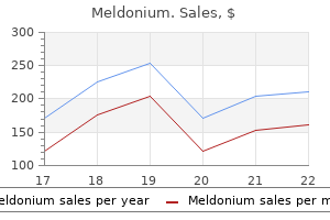
Meldonium
"Meldonium 500mg without prescription, medicine 6 times a day".
S. Milten, M.B.A., M.B.B.S., M.H.S.
Co-Director, The University of Arizona College of Medicine Phoenix
The outcomes of soft-tissue reconstruction typically merely change the arc of movement somewhat than normalize it xanthine medications purchase meldonium 250mg line. Postoperatively medications list a-z buy meldonium 250 mg low price, they typically have issue achieving full lively and passive flexion treatment hypercalcemia order 250 mg meldonium with amex. Oldfield (380) and Flatt (1) have stated that medications you can take during pregnancy order meldonium 250 mg amex, within the presence of marked radiographic evidence of bone and joint adjustments, corrective extension osteotomy may be most effective at bettering alignment and function. The published data about this salvage operation are too limited to allow goal evaluation (381). The articular floor can turn out to be incongruous, with notching of the bottom of the middle phalanx (372). This keeps the digital pulp of the affected fingers according to the opposite digital rays. However, because the contracture continues to progress past 30 levels and towards 90 degrees, it becomes harder for the affected person to compensate. Most clinicians recommend an initial treatment program of progressive passive extension and splinting with dynamic or progressive static splints. Parents are instructed to perform frequent home workout routines for their infants; affected adolescents are similarly instructed to perform a home program for themselves. Many clinicians (367, 373ͳ75) report success with a splinting program generally. Clinodactyly is irregular angulation (>10 degrees) of the digit in the radioulnar aircraft. Clinodactyly is also regularly related to syndromes (Holt-Oram, Turner, Silver, and Cornelia de Lange) and chromosomal abnormalities (trisomies 18 and 21), and will alert the primary neonatal examiner to look for related malformations or issues. For example, clinodactyly of the thumb is seen in Rubinstein-Taybi syndrome (386, 387) and diastrophic dwarfism (388, 389). Function is affected solely when the deformity is severe enough to impinge on the adjoining digit during flexion. In these rare situations, the progressive deformity is secondary to altered physeal development. Physeal bar resection and fats graft interposition have been reported by Vickers (390, 391) to restore longitudinal development and supply correction (392). Surgical realignment may be within the type of opening-wedge, closingwedge, and reverse-wedge osteotomies. The complications of osteotomy are persistent deformity and loss of interphalangeal movement. Loss of motion in a patient whose indication for surgical procedure was purely aesthetic is unacceptable to most sufferers, their households, and surgeons. Failure of separation happens with thumb index syndactylies, that are frequent with other syndactyly syndromes. Duplication is seen within the form of thumb polydactyly, undergrowth as thumb hypoplasia, and overgrowth as macrodactyly and triphalangeal thumbs. Also, thumb abnormalities are widespread as part of constriction band syndrome or generalized musculoskeletal disorders. The record is exhaustive and this part will cover the more common congenital thumb malformations. Less typically, remedy is by reconstruction, potentially including a microvascular toe metatarsophalangeal transfer to type a thumb carpometacarpal joint. The trigger seems to be a measurement mismatch between the flexor pollicis longus and the A1 pulley, resulting in progressive constriction. However, set off digits are seen with neurologic syndromes (trisomy 18) and mucopolysaccharidoses (119).
Placement of the pins in the distal fragment requires cautious localization of the fragment and avoidance of the extensor mechanism medications given during dialysis proven meldonium 250 mg. The fracture has generally been healing too long for profitable closed discount and percutaneous pinning to be carried out symptoms 6dpo generic meldonium 500 mg line. This fracture requires prompt attention medications heart disease purchase 500 mg meldonium, anatomic discount treatment variance cheap 250mg meldonium, and pin stabilization. If the fracture is left within the place proven here within the splint, there will be a problematic malunion. A pin is placed dorsally within the fracture site underneath fluoroscopic management and used for reducing the fracture. The subcondylar fossa can be reconstituted, and the fracture may be pinned percutaneously. Remodeling of the fracture is uncommon due to the significant distance from the physis, but it has been described in case reviews of each proximal and middle phalanx phalangeal neck fractures (462). Intercondylar fractures in younger children are often small osteochondral fractures. This is especially true in the middle phalanx if the harm is a crush damage that alters the local blood provide. The fracture is intraarticular, typically displaced, and requires anatomic discount for a profitable consequence. This injury requires acute anatomic reduction and pin stabilization to stop long-term loss of motion, malalignment, and potential ache and arthritis. The collateral ligament attachments to the fragment are preserved in order to reduce the danger of avascular necrosis. On event, bone grafting is critical for sustaining articular congruity and to stop collapse. Even with well-performed open discount, complications of this fracture can happen within the younger patient. Avascular necrosis often resolves by revascularization, however often not earlier than collapse. In the adolescent, therapy of intercondylar fractures is similar to that in adults. Anatomic discount and pin fixation are necessary to restore the joint floor and to forestall lack of reduction that can happen with this unstable fracture. Often, the procedure can be carried out closed, using distraction and a percutaneous towel clip to get hold of reduction (463). Restoration of an anatomic joint is the mandatory aim and will reduce the risk of lack of movement, malalignment, or long-term arthritis. Diaphysis-level phalangeal fractures are rare within the young baby and extra frequent within the teenager. However, shut inspection of the nail plates will reveal the digital malalignment. With passive wrist extension, the fingers flex and level toward the volar scaphoid tubercle. Tenodesis assessment ought to be carried out on all phalangeal and metacarpal fractures, regardless of radiographic appearance. If closed remedy is chosen, a finger should by no means be immobilized by itself, but should be secured to the adjoining digits to prevent subsequent lack of discount and malrotation. If the fracture is malrotated and unstable, reduction with pin or screw stabilization is important (463, 464). The malrotated digit impairs the function of the adjacent digits as a end result of the digits will overlap in flexion. Physeal fractures represent 30% to 70% of pediatric finger fractures (442ʹ44, 446).
Lengthening and deformity correction of the upper extremity by the Ilizarov approach symptoms multiple sclerosis purchase meldonium online from canada. P Preliminary experience with Ilizarov methodology in late reconstruction of radial hemimelia medicine hat jobs buy 500mg meldonium. Ulnar lengthening for unfavorable ulnar variance in hereditary multiple osteochondromas medicine 6 year in us order genuine meldonium line. Congenital forearm pseudarthrosis: report of six circumstances and evaluate of the literature treatment 3 antifungal generic meldonium 500mg otc. Congenital pseudarthrosis of the forearmδwo cases treated by free vascularized fibular graft. Use of free vascularized fibular graft for congenital ulnar pseudarthrosis: surgical determination making in the rising youngster. Congenital deformities of the upper extremities, Copenhagen, Denmark: Ejnar Munksgaard Forlag, 1950. Congenital amegakaryocytic thrombocytopenia and thrombocytopenia with absent radii. Absence of mutations in the HoxA10, HoxA11 and HoxD11 nucleotide coding sequences in thrombocytopenia with absent radius syndrome. A persevering with examine of sixty-eight patients with 100 and seventeen club palms. Microsurgical second toe-metatarsal bone switch for reconstructing congenital radial deficiency with hypoplastic thumb. Formation of radius congenitally absent, situation seven years after implantation of bone graft. Distraction lengthening and microvascular bone transplantation in the remedy of radial membership hand. Preoperative soft-tissue distraction for radial longitudinal deficiency: an analysis of indications and outcomes. Selective soft tissue launch preserves progress plate architecture throughout limb lengthening. Possible relationship between ulnar-mammary syndrome and cut up hand with aplasia of the ulna syndrome. Fine mapping of the autosomal dominant cut up hand/split foot locus on chromosome 7, band q21. Limb reduction defects in Emilia Romagna, Italy: epidemiological and genetic study in 173,109 consecutive births. A classification of cleft arms, based on clinical findings: principle of developmental mechanism. The use of a volar flap for restore of fingertip amputations: a preliminary report. Composite toe (phalanx and epiphysis) transfers in the reconstruction of the aphalangic hand. Proximal toe phalanx transplantation for bony stabilization and lengthening of partially aplastic digits. Distraction augmentation manoplasty: technique of lengthening digits or entire hands.
Congenital enlargement of the occipital condyles may have been the cause by increasing movement at this joint symptoms appendicitis discount meldonium 500 mg without prescription. Cervical radiculopathy and myelopathy in cerebral palsy (284Ͳ87) was first described within the athetoid types and subsequently within the spastic varieties medicine lodge ks order meldonium 250mg fast delivery. Athetoid cerebral palsy sufferers medicine misuse definition safe meldonium 250 mg, when compared with the normal inhabitants xerostomia medications that cause proven meldonium 250 mg, develop cervical disc degeneration at a younger age. Angular and listhetic instabilities are also more frequent and seem at a younger age (288). The mixture of disc degeneration and listhetic instability predisposes these patients to a relatively fast, progressive neurologic deficit. The signs are brachialgia and weakness of the higher extremity with decreased useful use or elevated paraparesis or tetraparesis (285Ͳ87). In ambulatory sufferers, a lack of ambulatory capability is often an indication of presentation. Atlantoaxial instability has been recently described in sufferers with extreme spastic quadriplegia; the symptoms are often apnea, opisthotonos, torticollis, respiratory problems, muscle tone abnormalities and hyperreflexia, and bradycardia (289). Flattening of the anterosuperior margins of the vertebral our bodies and beak-like projections of the anteroinferior margins are radiographic findings of the spondylosis. Myelography demonstrates stenosis, disc protrusion, osteophyte projection, and blocks in dye circulate, mostly at the C3-C4 and C4-C5 ranges. The kyphosis, herniated discs, and osteophytes result in nerve root and twine compression. It is believed that the exaggerated flexion and extension of the neck in these younger adults with cerebral palsy causes accelerated cervical degeneration and cervical stenosis earlier than in unaffected individuals, who develop stenosis in the late fourth and fifth many years of life. Exaggerated flexion and extension happens in sufferers with athetosis and writhing actions. Difficulty with head control can also trigger exaggerated flexion and extension within the spastic cerebral palsy affected person. Anterior discectomy, resection of osteophytes, and interbody fusion have been the best methods. However, postoperative immobilization could be a problem for some patients, and thus some authors also advocate a posterior wiring of the sides as properly to reduce the amount of time postoperative immobilization is needed (285). Posterior laminectomy alone (286) is contraindicated in cerebral palsy sufferers with developmental cervical stenosis as a result of this can increase the instability. A 14-year-old lady with spastic quadriparesis confirmed progressive loss of higher extremity perform with lack of capability to control her wheelchair and feed herself. A: the lateral radiograph reveals marked stenosis from C3-C6, as evidenced by a spinal canal-to-vertebral body ratio (Torg ratio) of <0. This stenosis was handled by posterior laminectomy from C3-C7 and posterior cervical fusion from C2-T1 utilizing Luque rectangle fixation with spinous process and facet wiring. It has been duplicated in animal fashions; a C3-C6 laminectomy in rising cats uniformly resulted in kyphosis; whereas regular cervical curves had been maintained in adult cats (300). The pure historical past of postlaminectomy kyphosis is unknown; however, the incidence of kyphosis when extensive cervical laminectomies are carried out in childhood varies from 33% to one hundred pc, with an total common of 70% (296). Postlaminectomy lordosis is much less frequent and is strongly correlated with a peak age at decompression of 4 years (296).
The sagittal view exhibits bounce alignment with gentle equinus on the ankle and vital flexion deformities at the hip and knee treatment works generic 500mg meldonium visa. The external foot progression angle is a mixture of exterior tibial torsion and pes valgus treatment thesaurus meldonium 500 mg discount. With the affected person within the susceptible place medicine yoga buy discount meldonium 250mg, a posteromedial incision medicine daughter order 250 mg meldonium with mastercard, 2 to 3 cm long, is made, centered over the musculotendinous junction of the gastrocnemius. The deep fascia is divided longitudinally, and the sural nerve and lesser saphenous vein are identified and guarded. The aircraft between the gastrocnemius and the soleus is identified from the medial aspect and developed by blunt dissection. Once the two layers have been separated, the aponeurosis of gastrocnemius is split transversely, the muscle bellies are allowed to recess proximally and are then sutured in the acceptable place (ankle in neutral, knee in extension). If the range of dorsiflexion remains to be limited to less than plantigrade, with the knee in extension, the fascia overlying the muscle belly of soleus could be divided transversely. Equinus leads to excessive loading of the forefoot and with time may trigger breaching of the midfoot. A collection of advanced segmental malalignments of the midfoot, hindfoot, and forefoot develops referred to as pes equinoplanovalgus, pes planoabductovalgus, or just "pes valgus. Symptoms could include pain and callosities over the collapsed medial arch, particularly the pinnacle of the talus. Cadaver dissection to show the distal gastrocnemius recession described by Strayer. The broad gastrocnemius aponeurosis has been divided transversely, on the distal extent of the medial gastrocnemius stomach. This ends in isolated lengthening of the gastrocnemius and is the most secure process for equinus in diplegia because it avoids the chance of weakening of the soleus. In some kids, lengthening of the soleus perhaps required and this is illustrated on the right where the soleus fascia has been divided transversely exposing the soleus muscle fibers in the intervening gap. A helpful guide to the radiographic useful anatomy of the foot, with regular values for a series of radiographic parameters, has been published (67). Factors affecting the selection of operative process embody the age of the patient and the scientific and radiographic severity of the deformity (133). The flexibility of the deformity is essential as a result of the generally used surgical techniques rely upon ligamentotaxis for the correction of all component parts of the deformity. The corrigibility of the deformity should be checked by putting the foot in an equinovarus position, whereas palpating the medial arch with special consideration to the talonavicular joint. As the foot moves into equinovarus, the medial arch ought to be restored and the navicular should cover the pinnacle of the talus. The midfoot can be stabilized and deformity corrected by lengthening of the lateral column of the foot (os calcis lengthening) or extra-articular fusion of the subtalar joint (170, 171, 184ͱ86). Os calcis lengthening corrects subtalar joint eversion and midfoot breaching by elongating the lateral column of the foot, driving the heel out of valgus, into relative varus and raising the medial arch. Arthrodesis of the subtalar joint is a dependable means of correcting hindfoot valgus and with secondary correction of the midfoot. A modified Fulford method is finest, with a cannulated screw passed via the talar neck, across the sinus tarsi into the calcaneum, mixed with iliac crest autograft or allograft (171). A fourth choice which is gaining in reputation is isolated fusion of the talo-navicular joint.